The acetabulum is an articular surface it articulates with the femoral head says stephen andy a.
Left acetabular roof.
The rest is formed by the pubis near the mid.
The angle is formed by a horizontal line connecting both triradiate cartilages hilgenreiner line and a second line which extends along the.
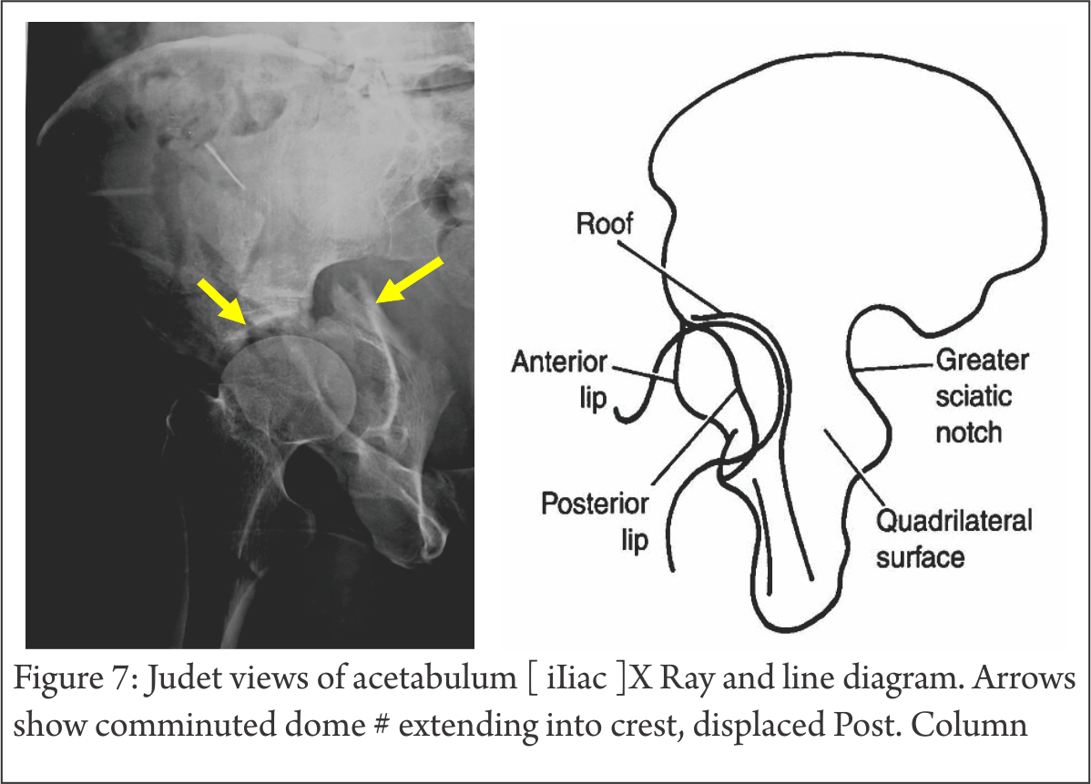
Acetabulum fractures can involve one or more of the two columns two walls or roof within the pelvis.
A benign cause of a lytic bone lesion is a solitary bone cyst as noted on the norwich image interpretation course.
Fractures occur in a bimodal distribution.
This occurs in people under 30 and typically appears in the long arm bone or the humerus.
The acetabular lines should be carefully scrutinized.
These hip socket fractures are not common they occur much less frequently than fractures of the upper femur or femoral head the ball portion of the joint.
It shows up on a medical imaging study such as an x ray as a point of increased density and is believed to be caused by overproduction of bone or cartilage cells.
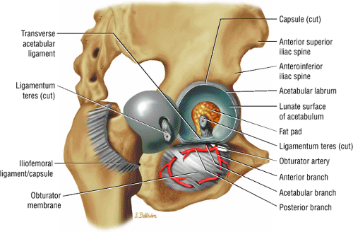
A deep semispherical socket cavity in the lateral surface of the hip with which the femoral head articulates located at the convergence of the ilium ischium and pubis.
The acetabulum is functionally comprised of two columns anterior and posterior and two walls anterior and posterior.
It is most useful in patients who have started to ossify the epiphysis since ossification diminishes the usefulness of ultrasound.
Sbcs were first discovered in 1940s but doctors are still uncertain about the reasons they form.
The acetabular angle is a radiographic measurement used when evaluating potential developmental dysplasia of the hip ddh.
The addition of oblique views of the affected hip may be necessary to evaluate the anterior and posterior columns of the acetabulum.
If the acetabulum is not well reduced or maligned or sometimes even if the surgeon is able to achieve the reduction and the alignment the cartilage coating the joints has been badly damaged and degenerates which is arthritis.
The majority of acetabular fractures are caused by some type of high energy event such as a car collision.
If an acetabular fracture is identified a ct scan is suggested to assess the position of the fracture fragments and to exclude intra articular loose bodies.
2012 farlex inc.
A bone island is a benign growth of bone or cartilage inside a bone usually within the marrow.
August 17 2020.
There are three bones of the os coxae hip bone that come together to form the acetabulum.
Sems m d an orthopedic trauma surgeon at mayo clinic s campus in rochester minnesota.
Mary mcmahon last modified date.
Sbcs occur in the subchondral bone which is the layer of bone right under cartilage.